Back Muscle Diagram / 10 Back Muscles Ideas Back Muscles Muscle Anatomy Shoulder Muscle Anatomy. National institute of arthritis and musculoskeletal and skin diseases: This muscular system diagram shows the major muscle groups from the back or posterior view. While muscles like the gluteals (in the thighs) are used any time we walk or climb a step, deep back muscles and abdominal muscles are usually not actively engaged during everyday activity. See back muscles and low back pain. Lower back muscle diagram anatomy.
To see a muscular system picture from the anterior (front) view click here. Archives of internal medicine, october 2011. Build wide lats with this back building exercise. The part of the nerve that emerges out of the spine is called the nerve root. Most of the time, back muscle pain is diagnosed then treated with little more than a prescription of rest, painkillers and muscle relaxants.
ሠBack Muscle Diagrams Labeled Stock Vectors Royalty Free Trapezius Illustrations Download On Depositphotos from st2.depositphotos.com Anatomynote.com found anatomy of back muscles diagram from plenty of anatomical pictures on the internet. Symptoms of muscle pain include: Facebook twitter google+ linkedin stumbleupon tumblr pinterest reddit vkontakte share via email print. While muscles like the gluteals (in the thighs) are used any time we walk or climb a step, deep back muscles and abdominal muscles are usually not actively engaged during everyday activity. Muscle spasms (contraction or stiffening of the back muscles) muscles that feel tight; This is a tutorial to quickly s. Five pairs of lumbar spinal nerves labeled l1 to l5 branch off your spinal cord and exit through small holes between the vertebrae. The back muscles represented on an anatomical chart and on a schematic view of the origin and insertion of the proper muscles of the back (iliocostal muscle of.
Does degenerative disc disease affect the lower back muscle?
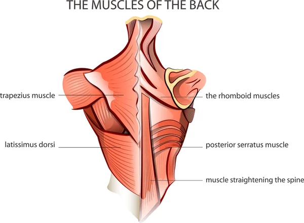
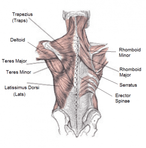
The back anatomy includes the latissimus dorsi, trapezius, erector spinae, rhomboid, and the teres major. These structures work together to support the body, enable a range of movements, and send messages from the brain to. Most of the time, back muscle pain is diagnosed then treated with little more than a prescription of rest, painkillers and muscle relaxants. How many muscles are in the back? The back has a total of 40 muscles. Below you'll see diagrams along with the names of the back muscles that may be the cause of your pain. Related posts of muscles of the lower back and buttocks diagram muscle anatomy study guide. They extend and rotate the head and neck. The human back extends from the buttocks to the posterior portion of the neck and shoulders. Facebook twitter google+ linkedin stumbleupon tumblr pinterest reddit vkontakte share via email print. The intermediate layer contains the erector spinae muscles, whose many functions include the extension and lateral flexion of the spine, head and neck. To see a muscular system picture from the anterior (front) view click here. The pelvis at the bottom of the back and the shoulders at the top of the back give the back.
They extend and rotate the head and neck. The back anatomy includes the latissimus dorsi, trapezius, erector spinae, rhomboid, and the teres major. The muscles of the back can be arranged into 3 categories based on their location: This is a diagram of the larger and more surface muscles of the low back. These are typical symptoms you might experience:
Torn Pulled Strained Back Muscles What You Didn T Know from cdn.shopify.com Below you'll see diagrams along with the names of the back muscles that may be the cause of your pain. This is a tutorial to quickly s. Related posts of back muscles chart muscle anatomy in shoulder. For example, some muscles located in the chest also help move the shoulders. Low back pain. sherman, k. Symptoms of muscle pain include: People with back pain people who experience headaches printing for use during doctor visits to communicate information about your symptoms quickly tracking your progress over time related tools: The intermediate layer contains the erector spinae muscles, whose many functions include the extension and lateral flexion of the spine, head and neck.
This muscular system diagram shows the major muscle groups from the back or posterior view.
The muscles of the back can be arranged into 3 categories based on their location: Your back hurting more when you move, less when you stay still; Related posts of muscles of the lower back and buttocks diagram muscle anatomy study guide. The most common type of back pain is muscle pain—also called muscle strain or soft tissue strain. Lower back muscle diagram anatomy. For example, some muscles located in the chest also help move the shoulders. We are pleased to provide you with the picture named anatomy of back muscles diagram.we hope this picture anatomy of back muscles diagram can help you study and research. The back has a total of 40 muscles. To see a muscular system picture from the anterior (front) view click here. Muscles of lower back diagram in this image, you will find an occipital bone, sternocleidomastoid, trapezius, deltoid in muscles of the lower back diagram. The back anatomy includes the latissimus dorsi, trapezius, erector spinae, rhomboid, and the teres major. Pain log more pain mapping tools Back muscle diagram back muscles big back big back muscles big lats bodybuilding secrets major back muscles.
While muscles like the gluteals (in the thighs) are used any time we walk or climb a step, deep back muscles and abdominal muscles are usually not actively engaged during everyday activity. The trapezius and latissimus dorsi muscles connect the upper limb to the vertebral column. Lower back muscle diagram anatomy. This is a tutorial to quickly s. The pelvis at the bottom of the back and the shoulders at the top of the back give the back.
The Complete Guide To Training Your Back from 9to5strength.com How many muscles are in the back? Back muscles, like any other muscle in the body, require adequate exercise to maintain strength and tone. Back to tracking tools main page. To learn more about the anatomy of the spine, watch this video. The intermediate layer contains the erector spinae muscles, whose many functions include the extension and lateral flexion of the spine, head and neck. The human back extends from the buttocks to the posterior portion of the neck and shoulders. The deep back muscles, also called intrinsic or true back muscles, consist of four layers of muscles: This picture also contains humerus, olecranon process of ulna, deep to tendon and so on.
The trapezius and latissimus dorsi muscles connect the upper limb to the vertebral column.
For more anatomy content please follow us and visit our website: The pelvis at the bottom of the back and the shoulders at the top of the back give the back. We are pleased to provide you with the picture named anatomy of back muscles diagram.we hope this picture anatomy of back muscles diagram can help you study and research. They extend and rotate the head and neck. Nerves in your lower back. Another common cause of lower back and hip pain is disc injury. A strained muscle in your lower back can be quite painful. Your back hurting more when you move, less when you stay still; Pain log more pain mapping tools This muscular system diagram shows the major muscle groups from the back or posterior view. The part of the nerve that emerges out of the spine is called the nerve root. Archives of internal medicine, october 2011. How many muscles are in the back?
Share :
Post a Comment
for "Back Muscle Diagram / 10 Back Muscles Ideas Back Muscles Muscle Anatomy Shoulder Muscle Anatomy"


Post a Comment for "Back Muscle Diagram / 10 Back Muscles Ideas Back Muscles Muscle Anatomy Shoulder Muscle Anatomy"